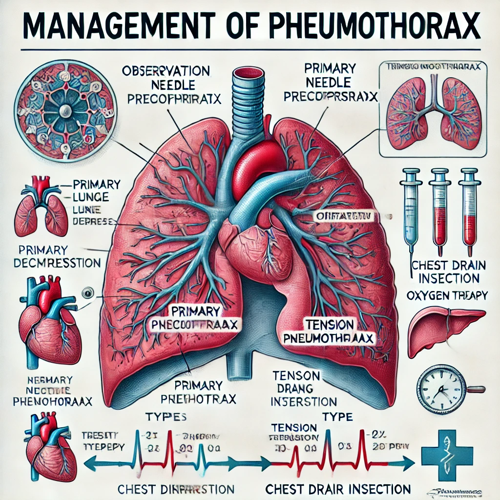
Pneumothorax refers to the presence of air in the pleural space, leading to lung collapse. It can be classified as spontaneous (primary or secondary) or traumatic. Management depends on the type, size, and clinical stability of the patient.
Types of Pneumothorax
- Primary Spontaneous Pneumothorax (PSP) – Occurs in healthy individuals, usually due to rupture of subpleural blebs.
- Secondary Spontaneous Pneumothorax (SSP) – Occurs in individuals with underlying lung disease (e.g., COPD, cystic fibrosis, tuberculosis).
- Traumatic Pneumothorax – Due to penetrating or blunt chest trauma.
- Iatrogenic Pneumothorax – Caused by medical procedures (e.g., central line insertion, lung biopsy, mechanical ventilation).
- Tension Pneumothorax – A life-threatening condition requiring immediate intervention.
Clinical Presentation
Symptoms:
- Sudden onset pleuritic chest pain
- Dyspnea (shortness of breath)
- Cough (often non-productive)
Signs:
- Reduced breath sounds on affected side
- Hyperresonance to percussion
- Decreased chest expansion
- Tracheal deviation (if tension pneumothorax)
Diagnosis
- Chest X-ray (CXR) – First-line investigation; shows air in pleural space with absence of lung markings.
- CT Scan – More sensitive for small pneumothoraces or complex cases.
- Ultrasound – Useful in emergency settings, detects absent lung sliding.
Management Based on Pneumothorax Type
1. Primary Spontaneous Pneumothorax (PSP)
- Small (<2 cm & asymptomatic): Observe and provide oxygen.
- Large (>2 cm or symptomatic):
- Aspirate with a 14-16G cannula or fine-bore chest drain.
- If aspiration fails, insert an intercostal chest drain (ICD) with underwater seal.
- Discharge once fully expanded with follow-up CXR.
2. Secondary Spontaneous Pneumothorax (SSP)
- If >50 years or significant dyspnea:
- >2 cm: Insert chest drain.
- 1-2 cm: Aspirate, if unsuccessful, insert a chest drain.
- <1 cm: Admit for observation and oxygen therapy.
3. Traumatic Pneumothorax
- Always insert a chest drain if lung injury is suspected.
- If associated with hemothorax (blood in pleural space), consider large-bore chest tube.
4. Tension Pneumothorax (Life-Threatening!)
- Immediate needle decompression at 2nd intercostal space, midclavicular line.
- Follow with definitive chest drain insertion.
- Do not wait for imaging if clinically suspected.
Chest Drain Insertion
- Use a safe triangle approach (5th intercostal space, midaxillary line).
- Secure with sutures and connect to an underwater seal drainage system.
- Monitor for re-expansion pulmonary edema, persistent leak, or infection.
Follow-Up & Prevention
- Smoking cessation (reduces recurrence risk in PSP).
- Pleurodesis for recurrent or persistent pneumothorax.
- Surgical options (VATS, pleurectomy) for severe cases.
Conclusion
Pneumothorax management is based on size, symptoms, and underlying lung disease. While primary cases may be observed, secondary, traumatic, and tension pneumothoraces require immediate intervention. Chest drain insertion remains the mainstay for moderate-to-severe cases, with further interventions for recurrent episodes.

