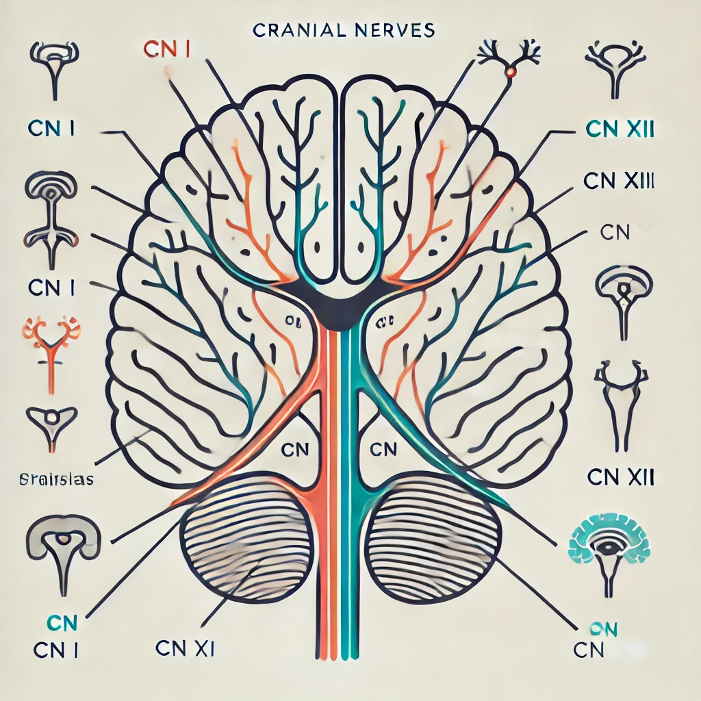
The cranial nerves (CN) are 12 pairs of nerves emerging from the brainstem, responsible for sensory, motor, and autonomic functions. Their lesions present with distinct clinical signs, making them a crucial topic in neurology and clinical practice.
1. Overview of Cranial Nerves
| Cranial Nerve | Function | Clinical Sign of Lesion |
|---|---|---|
| I. Olfactory (Sensory) | Smell | Anosmia (loss of smell) |
| II. Optic (Sensory) | Vision, pupillary reflex | Visual field loss, afferent pupillary defect |
| III. Oculomotor (Motor, Parasymp.) | Eye movement (except LR6, SO4), pupillary constriction | Ptosis, down & out eye, pupil dilation |
| IV. Trochlear (Motor) | Superior oblique (SO4) | Vertical diplopia, worsens on downward gaze |
| V. Trigeminal (Both) | Facial sensation, mastication, corneal reflex | Loss of sensation, weak bite, absent corneal reflex |
| VI. Abducens (Motor) | Lateral rectus (LR6) | Diplopia, inability to abduct eye |
| VII. Facial (Both, Parasymp.) | Facial expression, taste (ant. 2/3), salivation, lacrimation | Facial paralysis (UMN = spares forehead, LMN = whole side affected) |
| VIII. Vestibulocochlear (Sensory) | Hearing & balance | Hearing loss, vertigo, nystagmus |
| IX. Glossopharyngeal (Both, Parasymp.) | Taste (post. 1/3), gag reflex | Absent gag reflex, impaired taste |
| X. Vagus (Both, Parasymp.) | Swallowing, voice, parasympathetics | Dysphagia, uvula deviates opposite side |
| XI. Accessory (Motor) | SCM & trapezius | Shoulder droop, weak head turning |
| XII. Hypoglossal (Motor) | Tongue movement | Tongue deviates toward lesion |
2. Common Cranial Nerve Lesions and Clinical Presentation
(A) CN I – Olfactory Nerve Lesion
- Cause: Head trauma (cribriform plate fracture), neurodegenerative diseases (Parkinson’s, Alzheimer’s)
- Presentation: Anosmia (loss of smell), altered taste perception
(B) CN II – Optic Nerve Lesion
- Cause: Optic neuritis (MS), glaucoma, tumors
- Presentation:
- Monocular vision loss (optic nerve lesion)
- Bitemporal hemianopia (chiasm compression, e.g., pituitary tumor)
- Afferent pupillary defect (Marcus Gunn pupil)
(C) CN III – Oculomotor Nerve Lesion
- Cause: Posterior communicating artery aneurysm, uncal herniation
- Presentation:
- “Down and out” eye (loss of medial rectus, superior & inferior rectus)
- Ptosis (levator palpebrae weakness)
- Dilated pupil (mydriasis) (loss of parasympathetic function)
(D) CN IV – Trochlear Nerve Lesion
- Cause: Trauma, congenital
- Presentation:
- Vertical diplopia, worsens when looking downstairs
- Head tilt opposite side to compensate
(E) CN V – Trigeminal Nerve Lesion
- Cause: Trigeminal neuralgia, brainstem lesions
- Presentation:
- Loss of facial sensation (V1, V2, V3)
- Jaw deviation to side of lesion
- Absent corneal reflex
(F) CN VI – Abducens Nerve Lesion
- Cause: Increased ICP, cavernous sinus thrombosis
- Presentation:
- Horizontal diplopia
- Inability to abduct eye (unopposed medial rectus)
(G) CN VII – Facial Nerve Lesion
- Cause: Bell’s palsy (LMN), stroke (UMN)
- Presentation:
- LMN Lesion (Bell’s Palsy): Complete ipsilateral facial paralysis
- UMN Lesion (Stroke): Spares forehead, affects lower face
(H) CN VIII – Vestibulocochlear Nerve Lesion
- Cause: Acoustic neuroma, Meniere’s disease
- Presentation:
- Sensorineural hearing loss
- Vertigo, nystagmus
(I) CN IX – Glossopharyngeal Nerve Lesion
- Cause: Brainstem stroke, compression
- Presentation:
- Loss of gag reflex
- Impaired taste (posterior 1/3 of tongue)
(J) CN X – Vagus Nerve Lesion
- Cause: Brainstem stroke, surgery (thyroidectomy)
- Presentation:
- Dysphagia, hoarseness
- Uvula deviates opposite side
(K) CN XI – Accessory Nerve Lesion
- Cause: Neck trauma, surgery
- Presentation:
- Shoulder droop (trapezius paralysis)
- Weak head turning (SCM weakness)
(L) CN XII – Hypoglossal Nerve Lesion
- Cause: Medullary stroke, ALS
- Presentation:
- Tongue deviates toward lesion
3. Clinical Mnemonics for Cranial Nerves
(A) Names of the Cranial Nerves
Mnemonic: “Oh, Oh, Oh, To Touch And Feel Very Green Vegetables, AH!”
- Olfactory
- Optic
- Oculomotor
- Trochlear
- Trigeminal
- Abducens
- Facial
- Vestibulocochlear
- Glossopharyngeal
- Vagus
- Accessory
- Hypoglossal
(B) Cranial Nerve Function (Sensory, Motor, Both)
Mnemonic: “Some Say Money Matters, But My Brother Says Big Brains Matter More!”
- S = Sensory, M = Motor, B = Both
- I (S), II (S), III (M), IV (M), V (B), VI (M), VII (B), VIII (S), IX (B), X (B), XI (M), XII (M)
4. Diagnostic Approach
| Test | Indication |
|---|---|
| MRI Brain | Stroke, tumors, multiple cranial nerve lesions |
| CT Head | Trauma, hemorrhage |
| Electromyography (EMG) | Neuromuscular disorders (e.g., Bell’s palsy) |
| Audiometry | Hearing loss (CN VIII) |
| Fundoscopy | Optic nerve pathology (CN II) |
5. Key Takeaways
✅ Cranial nerves control vision, facial movement, swallowing, and autonomic functions.
✅ UMN lesions spare the forehead, LMN lesions affect the entire face (Bell’s Palsy).
✅ Horner’s syndrome (ptosis, miosis, anhidrosis) involves sympathetic dysfunction, not CN III.
✅ Stroke patients often present with CN IX and X deficits (dysphagia, uvula deviation).

