Congenital heart diseases (CHDs) are structural abnormalities of the heart present at birth. They range from simple, asymptomatic defects to severe, life-threatening conditions requiring urgent intervention. Advances in fetal screening, surgical correction, and catheter-based interventions have improved survival rates, allowing many CHD patients to reach adulthood.
2. Classification of Congenital Heart Diseases
CHDs are classified based on cyanosis, shunting patterns, and structural defects.
(A) Classification by Cyanosis
| Category |
Description |
Examples |
| Acyanotic CHDs (Left-to-Right Shunts) |
Increased pulmonary blood flow |
ASD, VSD, PDA, AVSD |
| Cyanotic CHDs (Right-to-Left Shunts) |
Deoxygenated blood enters systemic circulation |
Tetralogy of Fallot, Transposition of Great Arteries |
(B) Classification by Shunt Type
| Shunt Type |
Definition |
Examples |
| Left-to-Right Shunt |
Oxygenated blood recirculates to lungs |
ASD, VSD, PDA |
| Right-to-Left Shunt |
Deoxygenated blood enters systemic circulation |
Tetralogy of Fallot, Eisenmenger’s syndrome |
(C) Classification by Obstructive Defects
| Defect Type |
Description |
Examples |
| Outflow Tract Obstruction |
Narrowing of major vessels or valves |
Aortic stenosis, Coarctation of Aorta, Pulmonary stenosis |
3. Common Congenital Heart Diseases and Clinical Features
(A) Acyanotic Congenital Heart Diseases (Left-to-Right Shunts)
| Defect |
Pathophysiology |
Clinical Features |
Murmur |
| Atrial Septal Defect (ASD) |
Blood flows from LA → RA → RV |
Asymptomatic or dyspnea, paradoxical embolism |
Wide, fixed S2 splitting |
| Ventricular Septal Defect (VSD) |
Blood flows from LV → RV → Pulmonary circulation |
Most common CHD, loud murmur, CHF in large VSDs |
Harsh holosystolic murmur at LLSB |
| Patent Ductus Arteriosus (PDA) |
Persistent ductus arteriosus allows blood flow from Aorta → Pulmonary artery |
Continuous murmur, bounding pulses |
“Machine-like” continuous murmur |
| Atrioventricular Septal Defect (AVSD) |
Failure of endocardial cushion fusion, common in Down Syndrome |
CHF, failure to thrive, cyanosis in severe cases |
Systolic murmur + diastolic rumble |
(B) Cyanotic Congenital Heart Diseases (Right-to-Left Shunts)
| Defect |
Pathophysiology |
Clinical Features |
Murmur |
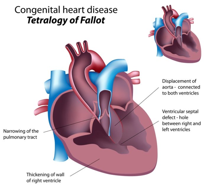
| Tetralogy of Fallot (TOF) |
RV outflow obstruction, VSD, Overriding aorta, RV hypertrophy |
Cyanosis, “Tet spells” (squatting improves symptoms) |
Harsh systolic ejection murmur at LUSB |
| Transposition of the Great Arteries (TGA) |
Aorta arises from RV, Pulmonary artery from LV |
Cyanosis at birth, requires PDA or VSD to survive |
Single loud S2 |
| Tricuspid Atresia |
Absence of tricuspid valve → No RV development |
Severe cyanosis, requires ASD or VSD for survival |
Holosystolic murmur (VSD) + Single S2 |
(C) Obstructive Congenital Heart Diseases
| Defect |
Pathophysiology |
Clinical Features |
Murmur |
| Coarctation of Aorta (CoA) |
Narrowing of the aortic arch, leads to LV hypertrophy |
Hypertension in upper limbs, weak lower limb pulses |
Systolic murmur radiating to back |
| Aortic Stenosis |
Narrowing of aortic valve |
Dyspnea, syncope, angina |
Ejection systolic murmur, crescendo-decrescendo |
| Pulmonary Stenosis |
Narrowing of pulmonary valve |
RV hypertrophy, exertional dyspnea |
Ejection systolic murmur at LUSB |
4. Diagnosis of Congenital Heart Diseases
| Investigation |
Purpose |
| Echocardiography (TTE/TEE) |
Gold standard for diagnosis |
| ECG |
May show RVH, LVH, axis deviation |
| Chest X-ray |
Cardiomegaly, increased pulmonary markings |
| Pulse Oximetry Screening |
Identifies cyanotic CHDs in newborns |
| Cardiac MRI/CT |
Detailed anatomical evaluation |
| Cardiac Catheterization |
Confirms complex CHDs and assesses pressures |
5. Management of Congenital Heart Diseases
(A) Medical Management
| Condition |
Treatment |
| ASD, Small VSD, PDA |
Observation, spontaneous closure possible |
| Large VSD, AVSD |
Diuretics, ACE inhibitors, surgical closure if symptomatic |
| PDA (Preterm Infants) |
Indomethacin/NSAIDs to close PDA |
| Cyanotic CHD (TOF, TGA, Tricuspid Atresia) |
Prostaglandin E1 (PGE1) to keep ductus open |
| Coarctation of Aorta |
IV Prostaglandin to maintain PDA until surgery |
(B) Surgical & Interventional Management
| Condition |
Procedure |
| ASD, VSD |
Surgical or catheter closure if large/symptomatic |
| TOF |
Complete repair (VSD closure, RVOT enlargement) |
| TGA |
Arterial switch operation in neonates |
| Coarctation of Aorta |
Balloon angioplasty or surgical repair |
| Severe Pulmonary Stenosis |
Balloon valvuloplasty |
6. Complications of Congenital Heart Diseases
🚨 Eisenmenger’s Syndrome → Pulmonary hypertension reverses shunt to right-to-left, causing cyanosis
🚨 Heart Failure → High-output failure in large left-to-right shunts
🚨 Arrhythmias → Common post-surgical repair
🚨 Endocarditis Risk → Prophylactic antibiotics needed in high-risk CHDs
7. Key Takeaways
✅ Left-to-right shunts (ASD, VSD, PDA) are acyanotic; right-to-left shunts (TOF, TGA) cause cyanosis.
✅ Tet spells in TOF improve with squatting.
✅ Echocardiography is the gold standard for diagnosis.
✅ Prostaglandin E1 keeps ductus open in ductal-dependent CHDs (TGA, CoA).
✅ Early surgical correction improves outcomes in critical CHDs.
Further Reading
- NHS Overview of Congenital Heart Disease: NHS UK
- European Society of Cardiology (ESC) Guidelines: ESC Guidelines

