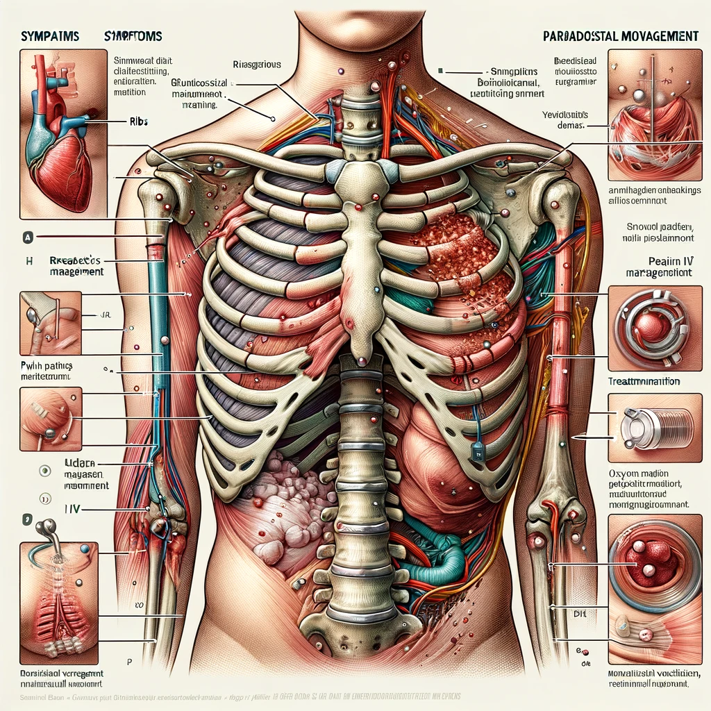
Flail chest is a life-threatening thoracic injury that occurs when a segment of the rib cage becomes detached from the rest of the chest wall due to multiple rib fractures. This results in paradoxical chest movement, impairing respiratory mechanics and leading to hypoxia.
Etiology and Causes
- Blunt chest trauma (e.g., motor vehicle accidents, falls, crush injuries)
- Severe impact sports injuries
- Penetrating trauma (less common but possible)
Clinical Presentation
Symptoms
- Severe chest pain and tenderness
- Dyspnea (difficulty breathing)
- Cyanosis (late sign of hypoxia)
- Shock (if associated with significant internal injuries)
Signs
- Paradoxical chest movement (affected segment moves inward during inspiration and outward during expiration)
- Tachypnea and respiratory distress
- Crepitus (palpable fractures)
- Decreased breath sounds (possible underlying lung contusion)
Diagnosis
1. Clinical Examination
- Inspect for paradoxical movement.
- Palpate for rib fractures and crepitus.
2. Imaging
A. Chest X-ray (CXR)
- Multiple rib fractures (≥3 ribs, in ≥2 places)
- Evidence of lung contusion or pneumothorax
B. CT Chest (Gold standard)
- Detects occult fractures and pulmonary contusions.
- Assesses for associated injuries (hemothorax, pneumothorax, pulmonary contusion).
C. Ultrasound (FAST Scan in Trauma Cases)
- Detects pneumothorax and hemothorax.
Management
1. Immediate Stabilization (ABCDE Approach)
A. Airway Management
- Ensure patency; intubate if severe respiratory failure.
- Consider early intubation and mechanical ventilation if oxygenation is compromised.
B. Breathing Support
- High-flow oxygen (target SpO₂ >94%).
- Non-invasive ventilation (NIV) or CPAP if mild-moderate respiratory distress.
- Mechanical ventilation if paradoxical movement impairs ventilation.
C. Circulation Management
- IV fluids (avoid excessive fluid resuscitation to prevent pulmonary edema).
- Monitor for hypotension and treat hemorrhagic shock if present.
2. Pain Management (Crucial for Preventing Respiratory Failure)
- Opioids (Morphine, Fentanyl) via PCA (Patient-Controlled Analgesia).
- Intercostal nerve blocks or epidural analgesia for severe cases.
- Avoid excessive sedation (to prevent hypoventilation and pneumonia).
3. Chest Wall Stabilization
- Surgical rib fixation (rib plating) is considered in severe cases or failure of conservative management.
- Indications for surgical fixation:
- Severe paradoxical movement with respiratory failure
- Recurrent pneumothorax
- Severe pain affecting ventilation
- Failure to wean from mechanical ventilation
4. Treat Associated Injuries
- Pneumothorax → Chest drain insertion.
- Hemothorax → Chest drain and fluid resuscitation.
- Pulmonary contusion → Oxygen therapy, NIV if mild-moderate, mechanical ventilation if severe.
5. Physiotherapy and Pulmonary Care
- Chest physiotherapy to prevent pneumonia and atelectasis.
- Incentive spirometry to encourage deep breathing.
- Early mobilization if feasible.
Complications
- Respiratory failure (due to inadequate ventilation)
- Pneumonia (due to poor lung expansion and secretion retention)
- Chronic pain and disability (from rib fractures)
- Long-term pulmonary dysfunction (in severe cases)
Prognosis and Follow-Up
- Mild cases (managed conservatively) have good recovery with pain control and physiotherapy.
- Severe cases (requiring ventilation or surgical fixation) have a prolonged hospital stay and require long-term pulmonary rehabilitation.
- Follow-up in respiratory clinic for persistent symptoms.
Conclusion
Flail chest is a serious condition requiring early recognition, respiratory support, effective pain management, and possibly surgical intervention. A multidisciplinary approach involving trauma, respiratory, and surgical teams improves outcomes and reduces complications. Adherence to UK trauma and respiratory guidelines (BTS, NICE, ATLS) ensures optimal patient care.

