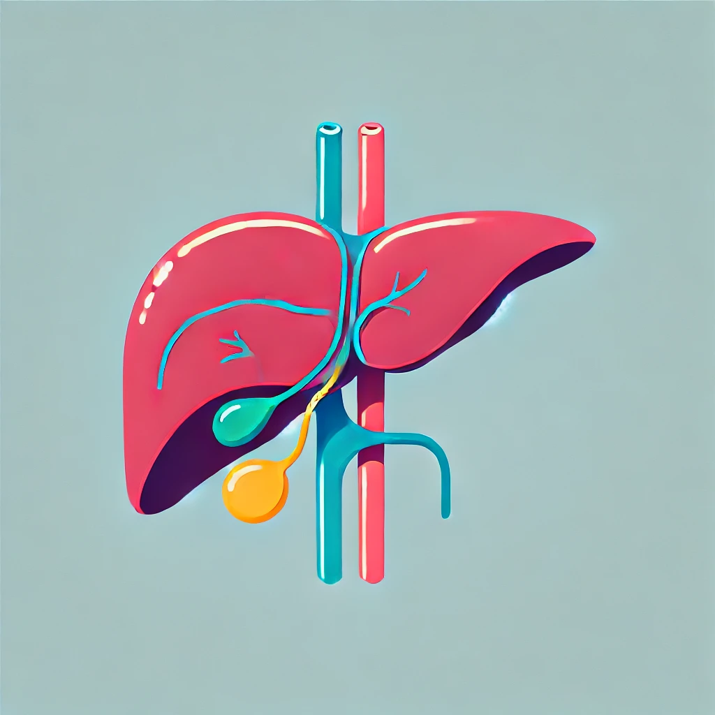
The hepatobiliary system plays a critical role in metabolism, detoxification, digestion, and waste elimination. It includes the liver, gallbladder, and bile ducts, forming a complex network essential for maintaining homeostasis. This article covers its anatomy, function, clinical significance, and diagnostic approach.
1. Anatomy of the Hepatobiliary System
The hepatobiliary system consists of:
- Liver – Largest gland, responsible for metabolism and detoxification.
- Gallbladder – Stores and concentrates bile.
- Biliary tree – Transports bile from the liver to the duodenum.
Liver Anatomy
- Lobes: Right and left lobes (divided by the falciform ligament).
- Microscopic structure: Contains hepatocytes organized into lobules with a central vein.
- Blood supply:
- Hepatic artery (25%) – Oxygenated blood from the celiac trunk.
- Portal vein (75%) – Nutrient-rich blood from the intestines.
Biliary Tree
| Structure | Function |
|---|---|
| Hepatocytes | Produce bile |
| Bile canaliculi | Collect bile from hepatocytes |
| Intrahepatic ducts | Merge to form hepatic ducts |
| Right & Left Hepatic Ducts | Drain bile from liver lobes |
| Common Hepatic Duct | Merges with cystic duct |
| Cystic Duct | Leads to gallbladder |
| Common Bile Duct | Drains bile into duodenum (via Ampulla of Vater) |
2. Functions of the Hepatobiliary System
| Function | Role |
|---|---|
| Bile Production | Aids in fat digestion |
| Detoxification | Removes drugs, toxins, and ammonia |
| Metabolism | Processes carbohydrates, fats, and proteins |
| Storage | Stores glycogen, vitamins (A, D, B12, K), and iron |
| Immune Function | Kupffer cells remove pathogens |
| Hormonal Regulation | Converts thyroid hormones, processes steroids |
3. Clinical Conditions of the Hepatobiliary System
(A) Hepatic Disorders
-
Hepatitis (Viral/Autoimmune/Toxic)
- Causes: Hepatitis A-E, alcohol, drugs (paracetamol overdose)
- Symptoms: Jaundice, hepatomegaly, fatigue, nausea
- Diagnostic Tests: LFTs, viral serology, ultrasound
- Management: Supportive care, antivirals (if indicated)
-
Cirrhosis & Liver Failure
- Causes: Chronic alcohol use, viral hepatitis, NAFLD
- Complications: Portal hypertension, ascites, hepatic encephalopathy
- Management: Lifestyle modifications, transplant for end-stage cases
-
Hepatocellular Carcinoma (HCC)
- Risk Factors: Cirrhosis, hepatitis B/C, aflatoxin exposure
- Diagnostic Tests: Alpha-fetoprotein (AFP), imaging (CT/MRI)
(B) Gallbladder & Biliary Tract Disorders
-
Cholelithiasis (Gallstones)
- Risk Factors: 5Fs (Female, Fat, Forty, Fertile, Fair-skinned)
- Symptoms: RUQ pain, nausea (post-fatty meal)
- Management: Cholecystectomy if symptomatic
-
Cholecystitis (Inflammation of the Gallbladder)
- Presentation: Murphy’s sign positive, fever, RUQ pain
- Investigations: Ultrasound (thickened gallbladder wall)
- Treatment: IV antibiotics, fluids, surgical removal
-
Choledocholithiasis & Cholangitis
- Charcot’s Triad (Cholangitis): Fever, jaundice, RUQ pain
- Management: ERCP (endoscopic stone removal)
4. Diagnostic Approach to Hepatobiliary Disease
| Test | Purpose |
|---|---|
| Liver Function Tests (LFTs) | Assess hepatocellular injury (ALT/AST), cholestasis (ALP/GGT) |
| Ultrasound | First-line for gallstones, liver pathology |
| CT/MRI | Evaluate liver masses, biliary obstructions |
| ERCP | Diagnostic & therapeutic for biliary obstruction |
| Liver Biopsy | Confirms cirrhosis, malignancy |
Liver Function Test Patterns
| LFT Abnormality | Possible Condition |
|---|---|
| ↑ ALT & AST | Hepatocellular damage (e.g., hepatitis) |
| ↑ ALP & GGT | Cholestasis (e.g., gallstones, PBC) |
| ↑ Bilirubin | Jaundice (pre/post-hepatic causes) |
5. Key Takeaways
✅ Hepatobiliary function is crucial for digestion, detoxification, and metabolism.
✅ Common conditions include hepatitis, cirrhosis, gallstones, and biliary obstruction.
✅ Diagnostic tools include LFTs, ultrasound, and ERCP for biliary diseases.
✅ Management varies from supportive care to surgical interventions like cholecystectomy or liver transplant.
Further Reading
- NHS Overview of Liver Diseases: NHS UK
- NICE Guidelines on Gallstones and Hepatitis: NICE Guidelines
Here’s a diagnostic flowchart for evaluating hepatobiliary disease, along with case-based scenarios to reinforce clinical decision-making.
1. Diagnostic Flowchart for Hepatobiliary Disease
This stepwise approach helps identify the underlying pathology based on symptoms, liver function test (LFT) patterns, and imaging findings.
                   Patient with suspected hepatobiliary disease
                                   │
               ┌───────────────────┴───────────────────┐
               │                                       │
     **Jaundice Present?**                   **RUQ Pain Present?**
               │                                       │
     ┌────────┴────────┐                    ┌────────┴─────────┐
     │                 │                    │                  │
**Conjugated**     **Unconjugated**   **Colicky Pain**     **Constant Pain**
     │                 │                    │                  │
**Obstructive**   **Hemolysis**       **Gallstones**      **Cholecystitis**
**Jaundice**      **(e.g., G6PD,**    **(Cholelithiasis)** **(Murphy’s sign +)**
**(ALP/GGT ↑,**   **Spherocytosis)**  │                   │
**Bilirubin ↑)**   **(LDH ↑, Hb ↓)**  │                   │
     │                                  │                   │
**Ultrasound → Dilated CBD?**   **Abdominal USS**   **Ultrasound**
     │                                  │                   │
**YES** → **ERCP for Obstruction** **Gallstones?** **Thick GB Wall?**
**NO** → **Liver Disease Workup**  **YES → Elective**  **YES → IV ABX,**
                                   **Cholecystectomy**  **Surgery**
2. Case-Based Clinical Scenarios
These scenarios highlight common hepatobiliary conditions and the stepwise diagnostic approach.
Case 1: Acute Hepatitis
Scenario:
A 30-year-old male presents with fatigue, nausea, RUQ pain, and jaundice. He recently traveled to an area endemic for hepatitis A. Examination shows mild hepatomegaly but no ascites or encephalopathy.
Approach:
- Initial Blood Work
- LFTs: ↑ ALT, ↑ AST, Bilirubin ↑, ALP normal
- Viral serology: Hepatitis A IgM positive
- Diagnosis: Acute Hepatitis A
- Management: Supportive care, hydration, avoid hepatotoxic drugs (e.g., paracetamol)
Case 2: Cirrhosis & Portal Hypertension
Scenario:
A 55-year-old male with a history of chronic alcohol use presents with abdominal distension, easy bruising, and confusion. Examination reveals spider naevi, caput medusae, shifting dullness, and asterixis.
Approach:
- Initial Blood Work
- LFTs: ↓ Albumin, ↑ Bilirubin, INR prolonged
- Ultrasound: Nodular liver, ascites, splenomegaly
- Endoscopy: Esophageal varices
- Diagnosis: Cirrhosis with portal hypertension
- Management:
- Ascites: Salt restriction, spironolactone, paracentesis
- Varices: Non-selective beta-blockers (propranolol), band ligation
- Encephalopathy: Lactulose, rifaximin
Case 3: Obstructive Jaundice (Choledocholithiasis)
Scenario:
A 45-year-old obese woman presents with jaundice, dark urine, pale stools, and pruritus. She reports colicky RUQ pain after fatty meals.
Approach:
- Initial Blood Work
- LFTs: ↑ ALP, ↑ GGT, ↑ Conjugated Bilirubin
- Abdominal ultrasound: Dilated common bile duct (CBD)
- Diagnosis: Choledocholithiasis (CBD stone)
- Management:
- ERCP for stone removal
- Laparoscopic cholecystectomy after ERCP
Case 4: Acute Cholecystitis
Scenario:
A 40-year-old woman presents with severe, persistent RUQ pain, fever, nausea, and vomiting. Examination reveals Murphy’s sign positive.
Approach:
- Initial Blood Work
- LFTs: Normal or mild ALP elevation
- WBC: ↑ Leukocytosis
- Ultrasound Findings:
- Gallbladder wall thickening, pericholecystic fluid, positive sonographic Murphy’s sign
- Diagnosis: Acute cholecystitis
- Management:
- IV antibiotics (ceftriaxone + metronidazole)
- Early laparoscopic cholecystectomy
Case 5: Acute Cholangitis (Life-Threatening)
Scenario:
A 60-year-old male presents with fever, jaundice, and RUQ pain (Charcot’s triad). He becomes hypotensive and confused (Reynolds’ pentad), suggesting sepsis.
Approach:
- Initial Blood Work
- LFTs: ↑ ALP, ↑ GGT, ↑ Bilirubin
- WBC: ↑ Leukocytosis
- Imaging: Dilated CBD on ultrasound
- Diagnosis: Acute ascending cholangitis
- Management:
- Urgent ERCP (biliary drainage)
- IV fluids, broad-spectrum IV antibiotics (piperacillin-tazobactam)
3. Key Takeaways for Clinical Practice
✅ Hepatobiliary diseases often present with jaundice, RUQ pain, or altered LFTs—tailor investigations accordingly.
✅ Use ultrasound as the first-line imaging for gallbladder and biliary diseases.
✅ ERCP is both diagnostic and therapeutic for biliary obstruction.
✅ Charcot’s triad (fever, jaundice, RUQ pain) is a medical emergency requiring immediate intervention.
✅ In cirrhosis, monitor for complications like variceal bleeding and hepatic encephalopathy.
Further Reading
- NHS Overview of Liver Diseases: NHS UK
- NICE Guidelines on Gallstones and Hepatitis: NICE Guidelines

