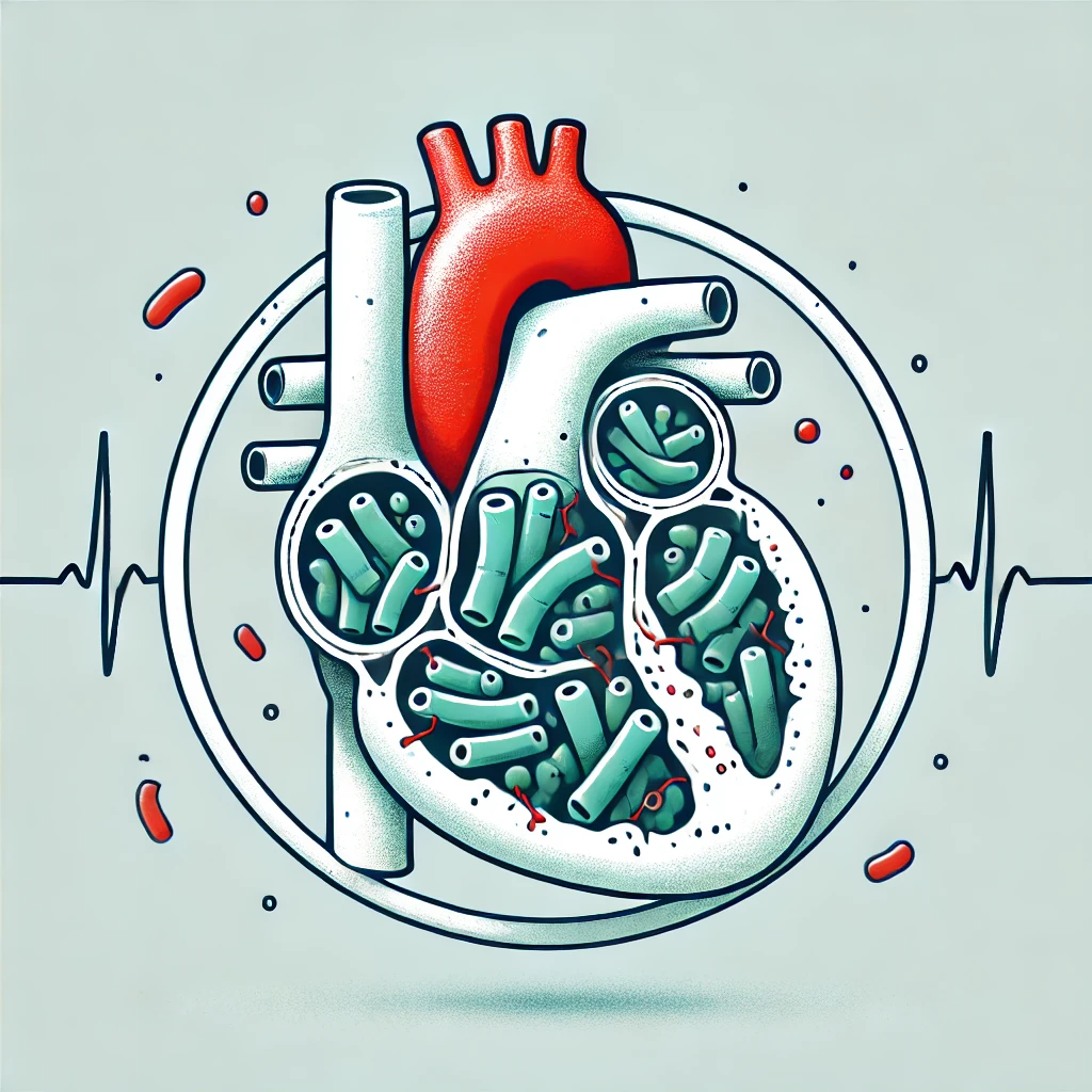
Infective Endocarditis (IE) is a life-threatening infection of the heart’s endocardium, most commonly affecting heart valves. It can lead to valvular destruction, heart failure, embolic events, and multi-organ complications. Early recognition and treatment are crucial to reduce morbidity and mortality.
2. Classification of Infective Endocarditis
| Type |
Description |
| Acute IE |
Rapid onset, highly virulent organisms (e.g., Staphylococcus aureus), aggressive progression |
| Subacute IE |
Indolent course, less virulent organisms (e.g., Streptococcus viridans), occurs on pre-existing valve disease |
| Native Valve Endocarditis (NVE) |
Infection on natural heart valves |
| Prosthetic Valve Endocarditis (PVE) |
Infection on prosthetic heart valves (early: <1 year post-surgery, late: >1 year post-surgery) |
| Intravenous Drug Use (IVDU) Endocarditis |
Commonly affects the tricuspid valve, caused by Staphylococcus aureus |
3. Risk Factors
| Category |
Risk Factors |
| Structural Heart Disease |
Rheumatic heart disease, bicuspid aortic valve, mitral valve prolapse |
| Prosthetic Material |
Prosthetic valves, indwelling cardiac devices (pacemakers, ICDs) |
| IV Drug Use |
Staphylococcus aureus is the most common causative organism |
| Immunosuppression |
HIV, chemotherapy, long-term corticosteroids |
| Recent Procedures |
Dental procedures, hemodialysis, intravascular catheters |
4. Pathophysiology
- Endothelial Injury → Turbulent blood flow (e.g., valvular disease) damages the endocardium.
- Platelet & Fibrin Deposition → Forms a non-bacterial thrombotic endocarditis (NBTE).
- Bacteremia → Organisms adhere to the thrombus, leading to vegetation formation.
- Embolization & Systemic Effects → Fragments of vegetation break off, causing stroke, infarcts, or septic emboli.
5. Clinical Features
(A) General Symptoms
| Symptoms |
Findings |
| Fever & Malaise |
Most common symptom (present in ~90%) |
| Night Sweats & Weight Loss |
Seen in subacute IE |
| New or Changing Murmur |
Indicates valvular involvement |
(B) Classic Signs of Infective Endocarditis
| Sign |
Description |
| Osler’s Nodes |
Painful, red nodules on fingertips/toes (immune complex deposition) |
| Janeway Lesions |
Non-tender macules on palms/soles (microemboli) |
| Splinter Hemorrhages |
Linear hemorrhages under fingernails |
| Roth Spots |
Retinal hemorrhages with pale centers |
| Clubbing |
Chronic IE cases |
| Splenomegaly |
More common in subacute IE |
(C) Embolic & Systemic Manifestations
| Complication |
Affected Organ |
Presentation |
| Stroke |
Brain |
Focal neurological deficits |
| Renal Infarct |
Kidneys |
Hematuria, flank pain |
| Septic Pulmonary Emboli |
Lungs (IVDU-associated IE) |
Respiratory symptoms, cavitary lesions on CXR |
| Mycotic Aneurysm |
Blood vessels |
Risk of rupture and hemorrhage |
6. Diagnosis of Infective Endocarditis
(A) Duke Criteria for IE Diagnosis
IE is diagnosed using the Modified Duke Criteria, which include major and minor criteria.
Major Criteria
- Positive Blood Cultures
- Staphylococcus aureus, Streptococcus viridans, Enterococcus (typical IE pathogens)
- Persistent bacteremia (≥2 positive blood cultures >12 hours apart)
- Endocardial Involvement (Echocardiography)
- Vegetation, abscess, or new dehiscence of prosthetic valve on transesophageal echocardiography (TEE)
Minor Criteria
- Predisposing Risk Factor (e.g., prosthetic valve, IVDU, heart disease)
- Fever >38°C
- Vascular Phenomena (e.g., Janeway lesions, embolic events)
- Immunologic Phenomena (e.g., Osler’s nodes, Roth spots)
- Microbiological Evidence (e.g., positive culture not meeting major criteria)
Definitive IE = 2 Major or 1 Major + 3 Minor or 5 Minor
Possible IE = 1 Major + 1 Minor or 3 Minor
(B) Laboratory & Imaging Workup
| Investigation |
Findings |
| Blood Cultures (3 sets before antibiotics) |
Bacteremia |
| Full Blood Count (FBC) |
Leukocytosis, anemia |
| Inflammatory Markers |
↑ ESR, CRP |
| Transesophageal Echocardiogram (TEE) |
Gold standard for detecting vegetations |
| ECG |
May show conduction abnormalities (suggests abscess) |
| CT/MRI |
To assess embolic complications |
7. Management of Infective Endocarditis
(A) Empirical Antibiotic Therapy (Before Culture Results)
| Suspected Organism |
First-Line Empirical Therapy |
| Native Valve (NVE) |
IV Vancomycin + IV Gentamicin |
| Prosthetic Valve (PVE) |
IV Vancomycin + IV Gentamicin + IV Rifampicin |
| IV Drug User (Tricuspid IE) |
IV Vancomycin |
(B) Targeted Antibiotic Therapy (After Culture Results)
| Causative Organism |
Antibiotic Regimen |
Duration |
| Staphylococcus aureus |
IV Flucloxacillin (MSSA) / IV Vancomycin (MRSA) |
4–6 weeks |
| Streptococcus viridans |
IV Benzylpenicillin ± Gentamicin |
4 weeks |
| Enterococcus spp. |
IV Ampicillin + Gentamicin |
4–6 weeks |
| HACEK Group |
IV Ceftriaxone |
4 weeks |
🚨 Prosthetic Valve IE requires ≥6 weeks of therapy
🚨 IV to oral switch is NOT recommended
(C) Indications for Surgery
| Indication |
Rationale |
| Severe Valve Dysfunction |
Refractory heart failure |
| Large Vegetations (>10 mm) |
High embolic risk |
| Persistent Infection |
Positive blood cultures despite antibiotics |
| Prosthetic Valve Endocarditis |
Higher risk of complications |
| Abscess Formation |
Conduction abnormalities (AV block on ECG) |
8. Prevention of Infective Endocarditis
(A) Who Needs Prophylaxis?
| High-Risk Group |
Examples |
| Prosthetic Valves |
Mechanical or bioprosthetic valves |
| Previous Infective Endocarditis |
History of IE |
| Congenital Heart Disease |
Unrepaired cyanotic defects, repaired defects with prosthetics |
(B) Antibiotic Prophylaxis for High-Risk Procedures
| Procedure |
Prophylactic Antibiotic |
| Dental (gingival manipulation) |
Amoxicillin 2g PO (or Clindamycin 600mg if allergic) |
| Respiratory (bronchoscopy with biopsy) |
Amoxicillin 2g IV |
| GI/GU Procedures |
Not routinely recommended |
9. Key Takeaways
✅ Fever + new murmur = Suspect IE!
✅ Duke Criteria guide diagnosis (TEE is the gold standard for vegetations).
✅ Blood cultures BEFORE antibiotics!
✅ Long-term IV antibiotics are required (4–6 weeks).
✅ Surgery indicated for severe valve damage, large vegetations, or abscesses.

