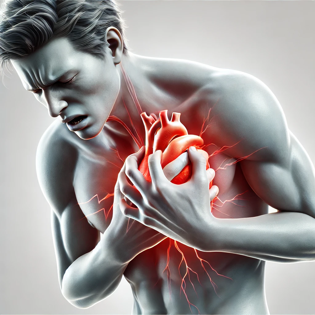
Myocardial infarction (MI), commonly known as a heart attack, occurs due to an acute obstruction of coronary blood flow, leading to ischemia and necrosis of myocardial tissue. It is a medical emergency requiring prompt recognition and intervention to reduce morbidity and mortality.
This article provides a comprehensive yet clinically practical approach to MI, covering pathophysiology, ECG changes, biomarkers, and guideline-based management strategies.
Further Reading: European Society of Cardiology (ESC) Guidelines on Myocardial Infarction
2. Pathophysiology of Myocardial Infarction
The pathophysiology of MI follows a series of events leading to irreversible myocardial necrosis:
- Plaque Rupture: Disruption of an atherosclerotic plaque within a coronary artery.
- Thrombus Formation: Platelet aggregation and fibrin deposition occlude the vessel.
- Myocardial Ischemia: Oxygen supply-demand mismatch leads to anaerobic metabolism.
- Tissue Necrosis: If ischemia persists, irreversible cell death occurs, triggering inflammation.
- Remodeling and Repair: Infarcted tissue is replaced by non-contractile fibrous scar tissue.
More on Atherosclerosis: American Heart Association (AHA) Explanation
3. Classification of Myocardial Infarction
(A) STEMI vs. NSTEMI
| Type of MI | ECG Findings | Pathophysiology |
|---|---|---|
| ST-Elevation Myocardial Infarction (STEMI) | ST-segment elevation in ≥2 contiguous leads | Complete occlusion of a coronary artery |
| Non-ST-Elevation Myocardial Infarction (NSTEMI) | ST depression, T wave inversions, no ST elevation | Partial occlusion or dynamic obstruction |
Clinical Importance: STEMI requires immediate reperfusion therapy, while NSTEMI is managed with risk stratification and possible delayed intervention.
(B) Universal Definition of MI (5 Types)
| Type | Cause |
|---|---|
| Type 1 | Atherosclerotic plaque rupture and thrombus formation |
| Type 2 | Supply-demand mismatch (e.g., sepsis, anemia, tachyarrhythmias) |
| Type 3 | Cardiac arrest with suspected MI but no biomarkers available |
| Type 4a | MI following percutaneous coronary intervention (PCI) |
| Type 5 | MI following coronary artery bypass grafting (CABG) |
Reference: Fourth Universal Definition of MI
4. Clinical Presentation
(A) Symptoms of MI
| Common Symptoms | Atypical Presentations |
|---|---|
| Chest Pain (retrosternal, crushing, radiates to left arm/jaw) | Silent MI (common in diabetics, elderly) |
| Dyspnea | Epigastric pain (may mimic gastritis) |
| Diaphoresis | Syncope |
| Nausea, Vomiting | Confusion (elderly) |
Key Clinical Pearl: Always consider silent MI in diabetic patients presenting with vague symptoms like fatigue or dizziness.
(B) Physical Examination
| Finding | Clinical Significance |
|---|---|
| Hypotension, cool extremities | Cardiogenic shock |
| Pulmonary crackles | Left ventricular failure |
| S4 Gallop | Ischemia-induced left ventricular stiffness |
Bedside Testing: Blood pressure, pulse oximetry, cardiac auscultation, and lung examination are essential for initial risk assessment.
5. ECG Changes in Myocardial Infarction
(A) ST-Segment Changes (Localizing Infarction)
| Infarct Location | Leads Affected | Coronary Artery |
|---|---|---|
| Anterior MI | V1-V4 | Left Anterior Descending (LAD) |
| Inferior MI | II, III, aVF | Right Coronary Artery (RCA) |
| Lateral MI | I, aVL, V5, V6 | Left Circumflex (LCx) |
| Posterior MI | ST depression in V1-V3 | RCA or LCx |
Clinical Pearl: Inferior MI may present with bradycardia due to RCA involvement affecting the AV node.
(B) Evolution of MI on ECG
| Phase | ECG Findings |
|---|---|
| Hyperacute Phase | Peaked T waves (minutes) |
| Acute Phase | ST elevation (hours) |
| Evolving Phase | Pathologic Q waves (days) |
| Chronic Phase | T wave inversions persist for weeks |
Guidelines on ECG Interpretation: NICE Guidelines on STEMI ECG Findings
6. Biomarkers for Diagnosis
(A) Cardiac Troponins (cTnI, cTnT)
- Most sensitive and specific marker for MI
- Rise within 2-4 hours, peak at 24-48 hours, remain elevated for 7-10 days
(B) Creatine Kinase-MB (CK-MB)
- Useful for detecting reinfarction (returns to baseline in 2-3 days)
(C) Other Markers
| Biomarker | Significance |
|---|---|
| Myoglobin | Early marker, but non-specific |
| BNP | Prognostic marker for heart failure in MI |
Reference for Biomarkers: American College of Cardiology (ACC) Recommendations
7. Management of Myocardial Infarction
(A) Initial Management (MONA-B)
| Treatment | Rationale |
|---|---|
| M – Morphine | Pain relief, reduces sympathetic activation |
| O – Oxygen | If SpO2 <90% |
| N – Nitrates | Vasodilation, symptom relief |
| A – Aspirin (300mg) | Antiplatelet therapy |
| B – Beta-blockers | Reduces myocardial oxygen demand |
(B) Reperfusion Therapy (STEMI)
| Treatment | Indication | Timing |
|---|---|---|
| Primary PCI | Preferred for STEMI | Within 90 minutes |
| Thrombolysis (tPA, Alteplase) | If PCI unavailable | Within 12 hours |
(C) Long-Term Secondary Prevention
| Medication | Benefit |
|---|---|
| Dual Antiplatelet Therapy (Aspirin + Clopidogrel) | Prevents further thrombotic events |
| Statins (Atorvastatin 80mg) | Stabilizes plaques, LDL reduction |
| ACE Inhibitors (Ramipril) | Reduces ventricular remodeling |
| Beta-blockers (Metoprolol, Bisoprolol) | Prevents arrhythmias, reduces mortality |
Post-MI Lifestyle Modifications: World Heart Federation Guidelines
8. Key Takeaways
✅ STEMI requires immediate reperfusion therapy (PCI or thrombolysis).
✅ NSTEMI is managed with risk stratification and medical therapy.
✅ Troponins are the gold-standard biomarkers for MI diagnosis.
✅ Dual antiplatelet therapy, statins, beta-blockers, and ACE inhibitors improve post-MI outcomes.

